Figure 1.
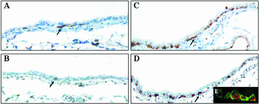
Proliferation and hyperplasia of basal cells in the bronchial epithelium of GCV-treated CCtk transgenic mice. CCtk mice were acutely exposed to vehicle (A and B) or 10 mg GCV (C and D), continuously exposed to BrdU during days 2 to 8 of the recovery period, and sacrificed on recovery day 10. Basal cells were detected by GSI-B4 lectin histochemistry (A and C) and S-phase cells were detected on adjacent serial sections by immunohistochemical detection of BrdU (C and D). Lectin and immune complexes were detected with diaminobenzidine (brown stain). Original magnification, ×400. Tissue sections from animals treated with GCV and recovered 3 days were analyzed by dual-immunofluorescence detection of GSI-B4 (green fluorescence) and BrdU (red fluorescence) and were analyzed by confocal microscopy (E). Original magnification, ×600.

