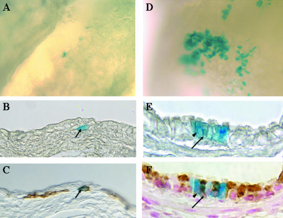
Figure 6.
Lineage of cytokeratin-14 expressing cells of the bronchial epithelium. K14-cre-ERt/RS bitransgenic mice were treated with naphthalene on day 0 and with Tamoxifen on recovery days 2 to 4. β-gal-expressing cells within the bronchial epithelium (blue-stained cells) were detected by wholemount histochemistry on recovery days 4 (A to C) and 20 (D to F). Tissue was initially imaged as a wholemount (A and D) and subsequently embedded, sectioned, and re-imaged (B and E). Sections containing β-gal-expressing cells were analyzed by immunohistochemistry for expression of K14 (C) or CCSP (F). Arrows in B and C indicate a β-gal/K14 dual-positive cell. Arrowheads in E and F indicate a β-gal-positive ciliated cell and the arrows indicate a β-gal/CCSP dual-positive cell.

