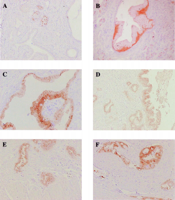
Figure 7.
Immunohistochemical analysis of S100P in normal pancreas, PanIN lesions, and PDAC. Representative images of S100P immunoreactivity in normal pancreatic islets (A), PanIN-1B (B), PanIN-2 (C), PanIN-3 (D), PDAC extending into the muscle wall (E), and perineural invasion (F). Original magnifications: ×100 (A, D, E); ×200 (B, C, F).

