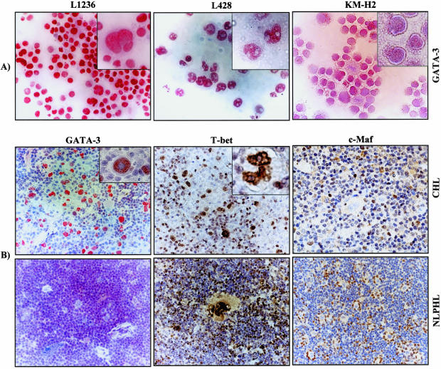
Figure 2.
Immunostaining of HL cell lines and tissues involved by HL for T-cell TFs. A: GATA-3 staining in HL cell lines. B: GATA-3, T-bet, c-Maf immunostaining of CHL and NLPHL cases. For GATA-3, frozen sections were used and for c-Maf and T-bet staining, paraffin sections were used. Original magnification ×100 for A–B, original magnification ×157.5 for insets.

