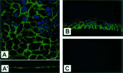
Figure 2.
Cell surface distribution of EMMPRIN in corneal epithelial cells in vitro. A: Confocal imaging of EMMPRIN staining in monolayer cultures of epithelial cells. Staining in A and vertical section in inset A′ show that EMMPRIN staining is distributed along the whole cell membrane and associated with small processes extending from the cells. B: EMMPRIN staining in stratified reconstituted epithelium in vitro shows a similar gradient to that seen in the central cornea. C: No signal was detected in control epithelium cultures when only the secondary antibody was used. Original magnifications, ×60.

