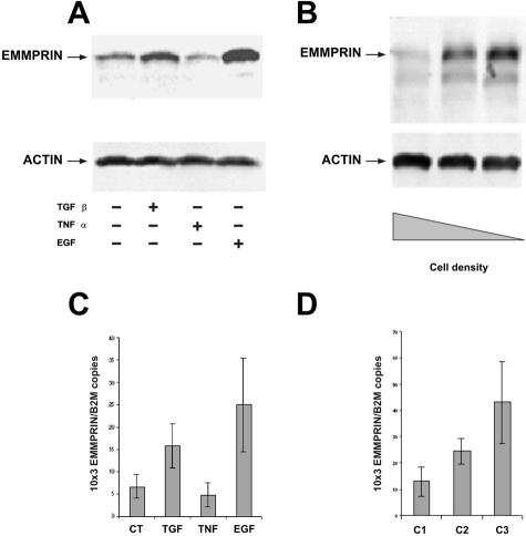
Figure 3.
Regulation of EMMPRIN in corneal epithelial cells by cytokines and cell density. Immunoblots (A, B) and real-time PCR (C, D) analysis of EMMPRIN regulation by cytokines (A, C) and cell density (B, D) in corneal epithelial cells. A and C: Cells were treated with TGF-β1 (10 ng/ml), EGF (10 ng/ml), or tumor necrosis factor-α (10 ng/ml) in serum-free medium for 24 hours (A) or 6 hours (C). B and D: Different cell density cultures were obtained by seeding 106 of cells in 25-cm2 (C1), 75-cm2 (C2), and 175-cm2 (C3) dishes. Cells were allowed to adhere for ∼24 hours to obtain confluent monolayer in the 25 cm2 (C1). For immunoblots, 20 μg of cell extracts were analyzed with either anti-EMMPRIN or anti-actin antibodies. C and D: Results are expressed as the mean of four separate experiments; bars, ±SD.

