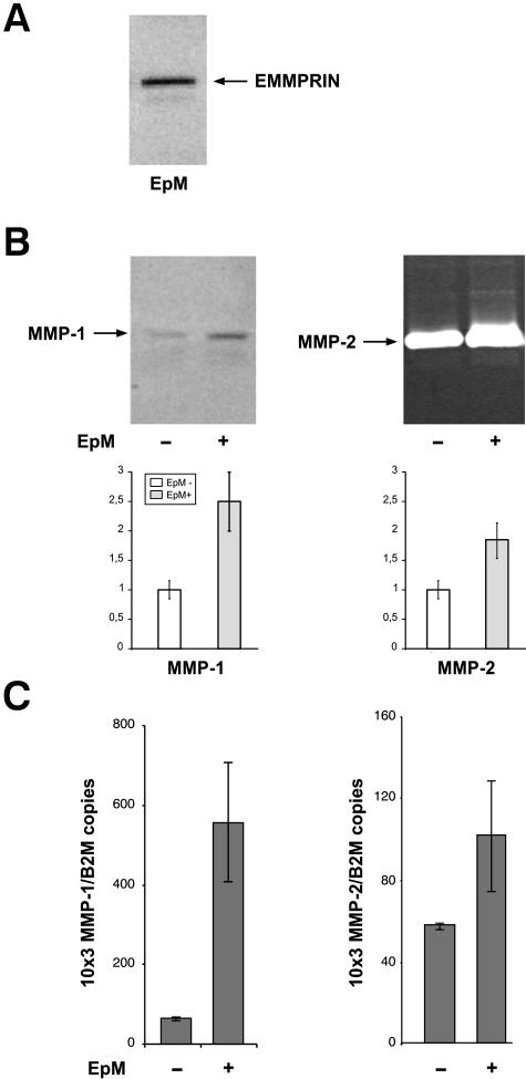
Figure 6.
EMMPRIN-containing corneal epithelial cell membranes stimulate MMP production in corneal fibroblasts in culture. A: Corneal epithelial cell membranes (EpM) were isolated by differential centrifugation and 10 μg of membrane extract were analyzed for EMMPRIN content by immunoblotting. One major band at 60 kd and a lower Mr faint band at ∼40 kd, corresponding to the high and low glycosylated forms of EMMPRIN, can be observed. B: Fibroblast cultures (80% confluent) were incubated with 20 μg/ml of EpM for 24 hours in serum-free medium after which the conditioned medium, 5× concentrated, was analyzed by immunoblot for MMP-1 and gelatin zymography for MMP-2. The amount of EpM added was derived from the same number of epithelial cells to that of fibroblasts. The graphs represent densitometry quantification of MMP-1 protein and MMP-2 gelatinolytic activity. C: Real-time PCR analysis of the same cultures as in B incubated with the membranes for 18 hours before RNA extraction. Results are expressed as the mean of triplicate experiments; bars, ±SD.

