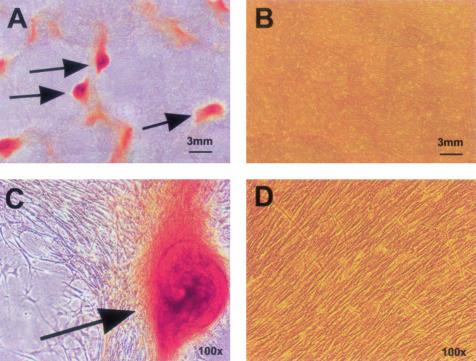
Figure 6.
Alizarin red staining of SAVICs after 7 days in culture on untreated type I bovine collagen (A and C), or on TGA-crosslinked collagen (B and D). Red stain indicates calcification. Calcific nodules (arrows) are macroscopic in size (A), while a uniform confluent culture of noncalcified cells is maintained, under the same conditions, but on TGA (B). Fields, ×100 (C and D) show details of cell morphology and staining.

