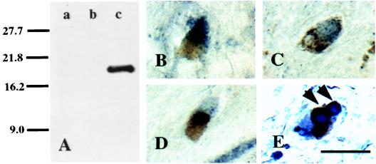
Figure 3.
Characterization of CM1 staining. (A) Specificity of the anti-CM1 polyclonal rabbit antibody. Western immunoblot of activated caspase-3 from 50 μg of SNpc protein extracted from four parkinsonian mesencephalons (lane a), 15 μg of untreated Jurkat cell lysate (lane b), and 15 μg of Jurkat cell lysate preincubated with 500 ng of human recombinant caspase-3 for 60 min at 37°C (lane c) after SDS/PAGE. Molecular mass markers in the first lane are given in kDa and allow identification of the cleaved p17 subunit at molecular masses of 17 kDa in lane c (A). Note that in the homogenate of the parkinsonian SNpc, activated caspase-3 is not detectable because of its presence in only a few cells. (B–D) High-power photomicrograph showing cytosolic-activated caspase-3 immunostaining (CM1 antibody) of SNpc neuromelanin-containing neurons in transverse sections of control SNpc. (E) High-power photomicrograph showing Lewy bodies stained with antiubiquitin (arrow) and co-CM1 immunostaining of SNpc neuromelanin-containing neurons in transverse sections of PD SNpc. Note the presence of both Lewy bodies and CM1 staining in a single neuron. (Bar = 30 μm.)

