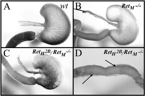
Figure 6.
Acetylcholinesterase staining of ENS in the stomach and duodenum. A: Wild-type stomach and duodenum (arrow) showing the fine network of nerves of the ENS. B: Stomach and duodenum (arrow) of mouse lacking Ret (genotype RetM−/−) showing aganglionosis of the ENS. C: Stomach and duodenum (arrow) of transgenic mouse with genotype RetH2B/RetM−/− showing partial rescue of the ENS by expression of human RetH2B. D: Distal to the stomach in the RetH2B/RetM−/− mouse, acetylcholinesterase staining is limited to a small number of longitudinal ENS bundles (arrows) in the proximal small intestine.

