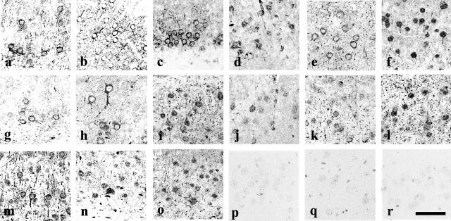
Figure 3.
Phosphorylation of accumulated tau. In the Tg mice brains, anti-tau154 antibody-labeled tau accumulations are seen in neurons and processes and as small round dot-like stains (a). These structures are labeled by both E1 (b) and PHF-1 antibodies (c). Antibody CP13 (d), Alz-50 (e) labeled neuronal cell bodies and dendrites. Anti-ubiquitin antibody stained neuronal cell bodies and dendrites (f). Phosphorylated tau antibodies PS199, AT8 and PT205, PT231/PS235, PS396, PS413 labeled neuronal cell bodies and dendrites (g–l). Anti-GSK-3β antibody labeled cell bodies of neurons (m). Anti-PY216 antibody against activated GSK-3β immunostained cell bodies of neurons (n), which are also stained by Cdk5 (o). Neither anti-PS9 against non-activated GSK-3β, anti-GSK-3α, nor anti-MAPK labeled these neurons (p to r). Bar, 50 μm.

