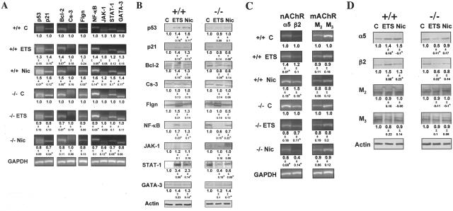
Figure 2.
Null mutation of α3 nAChR subunit abolishes ETS- and Nic-dependent changes in KC gene expression in in vivo experiments. To determine the role of α3 nAChRs in mediating effects of ETS and pure Nic on KCs, the gene expression at the mRNA and protein levels was analyzed using RT-PCR and WB assays, respectively. Asterisks indicate significant (P < 0.05) differences from control. A and C: RT-PCR analysis of ETS and Nic effects on gene expression in oral mucosa of exposed α3−/− mice. Gene-specific RT-PCR primers were designed to amplify the murine p53, p21, Bcl-2, Cs-3, Flgn, NF-κB, JAK-1, STAT-1, and GATA-3 (A), or α3 and β2 nAChR subunit, or M2 and M3 mAChR subtype (C) genes (Table 2). The ratio data underneath the bands are the means ± SD of the values obtained in at least three independent experiments. The images show representative bands in gels. B and D: WB analysis of ETS and Nic effects on gene expression in oral mucosa of exposed α3−/− mice. The proteins levels of p53, p21, Bcl-2, Cs-3, Flgn, NF-κB, JAK-1, STAT-1, and GATA-3 (B), or α3 and β2 nAChR subunits, or M2 and M3 mAChR subtypes (D) were analyzed by WB of total protein isolated from the same α3+/+ and α3−/− mice described in B and D, and analyzed by WB as detailed in the legend to Figure 1, C and E, using primary antibodies listed in Table 4. The gene expression ratio of 1 was assigned to oral tissue samples from α3+/+ mice. The ratio data underneath the bands are the means ± SD of the values obtained in at least three independent experiments. The images show typical bands in gels.

