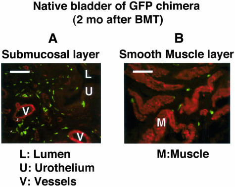
Figure 1.
Bladder of bone marrow-chimeric rat and the distribution of transplanted marrow-derived cells in the native bladder. Two months after bone marrow transplantation, the bladder wall was excised at the surgery and was stained for αSMA (red) and GFP (green). A: Submucosal area: relatively higher numbers of transplanted GFP-positive cells, which are negative for αSMA, are distributed. B: Smooth muscle layer: fewer cells are located at the periphery of smooth muscle fascicles. Bar, 100 μm (magnification, ×400). After fixation in 4% paraformaldehyde for 3 hours at 4°C, the specimen was processed in a serial sucrose gradient, frozen and embedded in cryocompound. Frozen sections (5-μm thick) were stained with hematoxylin and eosin (H&E) or double-immunostained with rabbit anti-GFP antibody (1:200) and mouse anti-αSMA antibody (1:100), and then visualized following incubation with fluorescein-conjugated anti-rabbit immunoglobulin and Texas-red-conjugated anti-mouse immunoglobulin.

