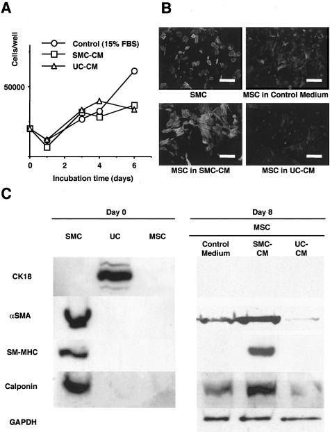
Figure 4.
Marrow cells take distinct phenotype in conditioned media of bladder cells. A: Growth curve of MSC cultured in SMC-CM, UC-CM, and control medium. MSC proliferated at equivalent rate in SMC-CM and UC-CM. B: Immunofluorescence for αSMA in cultured SMC and MSC cultured in SMC-CM, UC-CM, and control medium (magnification, ×400). The cells were fixed with 4% paraformaldehyde, incubated with αSMA antibody (1:100), and visualized with fluorescein-isothiocyanate (FITC)-conjugated anti-mouse immunoglobulin. The nucleus was counterstained with Hoechst 33258 dye. C: Left: Cultured SMC expressed αSMA, SM-MHC and calponin. UC expressed cytokeratin. MSC expressed little or no αSMA and no other SMC markers. Right: MSC cultured in SMC-CM expressed stronger expression of all three SMC markers than MSC cultured in control medium or in UC-CM.

