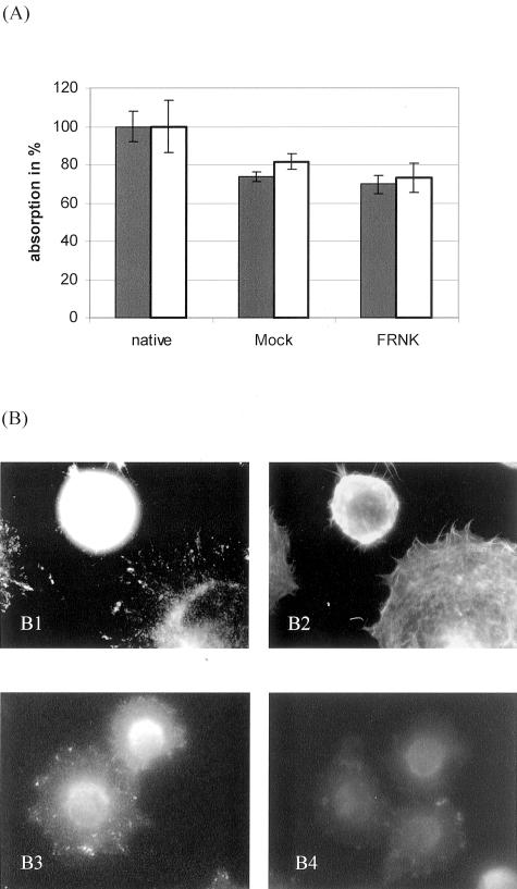
Figure 3.
Static cell adhesion and spreading of FRNK-transfected cells. A: Hep-G2 cells transfected with EGFP-plasmid (//) or pcDNA3.1-plasmid () containing FRNK or in its genuine form (mock) were allowed to adhere to collagen IV in microtiter plate assays for 30 minutes. Significant differences in the numbers of adherent cells were not found between FRNK- and control-transfected cells. Values are shown as mean ± SD from quadruplicate experiments. Comparable results were obtained using collagen I or colon carcinoma cells (not shown). B: Hep-G2 cells adherent to collagen IV for 30 minutes were fluorescence-stained for FAK (epitope within FRNK-fragment, B1) and F-actin (phalloidin, B2) in a double-labeling technique. The cell expressing FRNK remained round without cell spreading and reorganization of the actin cytoskeleton. In addition, FRNK-transfected cells did not show adhesion-specific phosphorylation of FAK (B4), whereas mock-transfected cells demonstrated typical focal adhesions with phosphorylated Tyr397 residues of FAK (B3). This inhibition was confirmed by Western blot (not shown).

