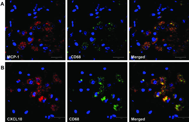
Figure 4.
A representative image of co-localization of MCP-1/CXCL10 and macrophages in the lungs of SHIV-infected macaques with pneumonia. Goat anti-MCP-1 polyclonal antibody (A) or mouse anti-CXCL10 monoclonal antibody (B) was used as primary antibody to stain the lung sections, followed by treatment with Alexa Fluor 594-conjugated secondary antibody. Macrophage-specific FITC-tagged mouse anti-CD68 antibody was then used as second primary antibody. After the final washing, the slides were mounted in SlowFade antifade reagent (with DAPI, blue stain) and images were captured by confocal microscopy.

