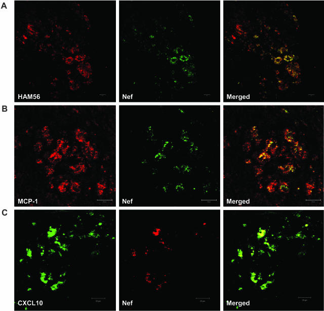
Figure 5.
A: A representative image of co-localization of SHIV and macrophages in the lungs of SHIV-infected macaque with pneumonia. Lung sections were first stained with rabbit anti-Nef polyclonal antibody, followed by Alexa Fluor 488-conjugated anti-rabbit secondary antibody; then the sections were stained with mouse anti-HAM56 monoclonal antibody, followed by Alexa Fluor 594-conjugated anti-mouse secondary antibody. B and C: Co-localization of MCP-1/CXCL10 with SHIV-infected cells in the lungs of macaque PJX. Goat anti-MCP-1 polyclonal antibody (B) or mouse anti-CXCL10 monoclonal antibody (C) and secondary antibody (Alexa Fluor 594-conjugated donkey anti-goat secondary antibody for MCP-1 or Alexa Fluor 488-conjugated donkey anti-mouse secondary antibody for CXCL10) were used to stain the lung sections from macaque PJX, The sections were then stained with rabbit anti-Nef polyclonal antibody, followed by Alexa Fluor 488-conjugated (staining MCP-1) or Alexa Fluor 594-conjugated (staining CXCL10) donkey anti-rabbit antibody as the second secondary antibody. After the final washing, the slides were mounted in SlowFade antifade reagent and images were captured by confocal microscopy.

