Figure 5.
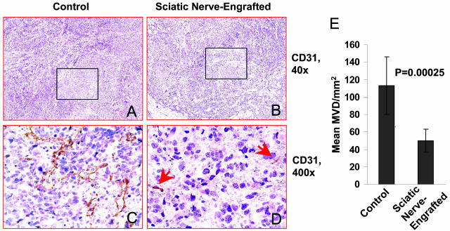
Representative photographs taken from CD31-immunostained slides of tumors engrafted outside (A and C) versus inside the sciatic nerve (B and D). Arrows in D point to smaller vessels seen in the sciatic nerve-engrafted tumor. E: The MVDs of the sciatic nerve-engrafted tumors and controls are shown in the bar graph.

