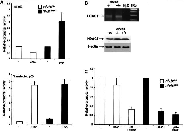
Figure 10.
HDAC1-mediated repression of TNF-α transcription is p50-dependent. A (top): Activated HSCs were transfected with 10 ng of pRLTK and 1 μg of pTNF-α-Luc for 24 hours before 24 hours of treatment with or without 500 nmol/L TSA. Luciferase activities were determined, normalized to pRLTK activity, and expressed as promoter activities relative to activity measured in cells lacking TSA treatment. A (bottom): As above but with the inclusion of pCDNA3p50 in the transfection mixture. All transfections were performed in triplicate. B (top): Total RNA was isolated from nfκb1−/− and nfκb1+/+ HSCs and used as a template for RT-PCR detection of HDAC1. B (bottom): Whole cell extracts were isolated from nfκb1−/−- and nfκb1+/+-activated HSCs and 25 μg of protein was used to immunoblot for HDAC1 and β-actin. C: Activated HSCs were transfected with 10 ng of pRLTK and 0.5 μg of pTNF-α-Luc alone (−) or together with 1 μg of pCDNA3 and 2 μg of pHDAC1 (HDAC1) or 1 μg of pCDNA3-p50 and 2 μg of pHDAC1 (p50 + HDAC1). After 48 hours, luciferase activities were determined, normalized to pRLTK activity, and expressed as promoter activities relative to activity in cells transfected with pTNF-α-Luc alone (mean ± SE of triplicate transfections).

