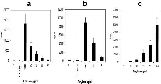
Figure 1.
DA(RP) (a) or Lewis (b) anti-amylase T cell lines were evaluated for antigen specificity in a proliferation assay using syngeneic antigen-presenting cells and varying concentrations of antigen. c: DA(RP), RT1.AaB/Dl, were stimulated with varying concentrations of amylase in the presence of Lewis, RT1.AlB/Dl, antigen-presenting cells to confirm MHC II restriction of the T cells. Vertical axes represent 3H-thymidine incorporation in dividing cells. Results are expressed as cpm ± SD.

