Figure 3.
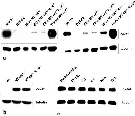
a: Analysis of ret expression in the skin of adult MT-ret+/− mice by two independent immunoblots. Ret protein levels are rather low in the skin of MT-ret+/− mice, and there is no detectable decrease in ret expression in mice lacking one (IL-6+/−) or both (IL-6−/−) copies of the IL-6 gene. Melanomas of an MT-ret+/− and an MT-ret+/−/IL-6+/− mouse express larger amounts of ret protein, as does the Mel25 cell line. As expected, the B16-F0 cell line does not show any ret expression. b: Immunoblot analysis of MT-ret+/− melanomas. Tumors derived from IL-6+/+ and IL-6−/− mice show similar ret protein expression. Wt: skin from wild-type BL/6 mouse. c: No increase in ret protein expression in immunoblots of Mel25 cells after stimulation with H-IL-6 for 15 minutes to 72 hours.

