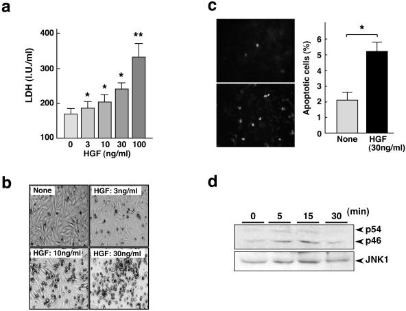
Figure 4.
Apoptosis and JNK1 phosphorylation promoted by HGF in portal myofibroblasts. a: Change in LDH activity in culture media. LDH activity was measured in conditioned media obtained after cultivation of portal myofibroblasts for 48 hours in the absence or presence of HGF. Each value represents the mean ± SD (n = 6). *, P < 0.05 and **, P < 0.01 versus nontreated control cells. b: Appearances of portal myofibroblasts cultured in the absence or presence of 3 ng/ml, 10 ng/ml, and 30 ng/ml HGF for 5 days. c: Apoptosis in activated liver myofibroblasts as detected by TUNEL staining. Cells were cultured in the absence or presence of HGF for 48 hours and subjected to TUNEL staining (left). The number of TUNEL-positive cells is shown at the right. Each value represents the mean ± SD. *, P < 0.01. d: Phosphorylation of JNK-1 enhanced by HGF in portal myofibroblasts. Phosphorylation of two forms of JNK-1 (p54 and p46) was detected using Western blots.

