Figure 2.
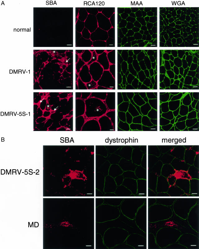
Lectin histochemical analyses of biopsied skeletal muscle tissues. A: Transverse cryosections of muscle biopsy specimens from normal controls (N1 or N2) and DMRV patients (DMRV-1 and DMRV-5S-2) were stained with rhodamine isothiocyanate-labeled SBA, RCA120, and fluorescein isothiocyanate-labeled MAA and WGA. Increased reactivity for SBA was observed mainly in the connective tissue, the rimmed vacuoles (asterisks), and the cytoplasm of the DMRV atrophic fibers (arrows). RCA120 reactivity was found in the sarcolemmal region, the connective tissue, and the rimmed vacuoles (asterisks) of the DMRV skeletal muscles. B: Transverse cryosections of muscle biopsy specimens from a DMRV patient (DMRV-5S-2) and a MD patient were co-stained with SBA lectin and an antibody against dystrophin. The merged images show that the SBA reactivity was not co-localized with dystrophin, indicating that the SBA-reactive materials were not co-localized with the sarcolemma in the DMRV and MD patients. The most intense SBA reactivity was found in the cytoplasm around rimmed vacuoles of the DMRV patient. SBA reactivity was also found in the connective tissues of both the DMRV and MD patients. Scale bars: 50 μm (A: SBA, MAA, and WGA); 20 μm (A: RCA120, B).

