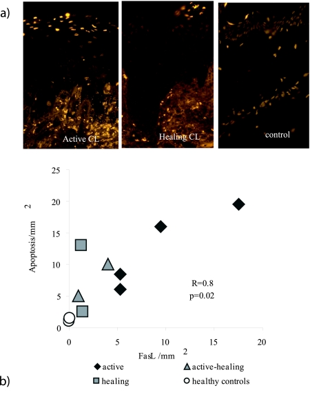
Figure 2.
Apoptotic cells were present in epidermis and correlated with the number of FasL-expressing cells found in dermis. a: Apoptosis was visualized by staining for TUNEL-positive cells (TMR) in paraffin-embedded sections. Apoptotic cells are seen as bright yellow. More apoptosis was measured in active lesions as compared to healthy or controls. Nonapoptotic nuclei are seen as orange-brown. b: The occurrence of FasL-expressing cells in inflammatory areas in dermis correlated with the occurrence of apoptotic cells in epidermis in CL biopsies (r = 0.8, P < 0.02, n = 8).

