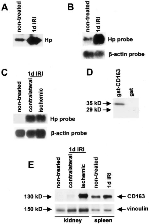
Figure 4.
Hp expression after IRI. A: Western blotting of plasma proteins of a wild-type mouse before and one day after IRI assayed with an anti-Hp antibody. B: Northern blotting of total RNA extracted from the liver of a non-treated wild-type mouse and of a mouse subjected to IRI one day after surgery, sequentially analyzed with 32P-dCTP-labeled probes to Hp and β-actin. C: Northern blotting of poly-A+ RNA extracted from the kidney of a non-treated wild-type mouse and from the contralateral and ischemic kidney of a wild-type mouse subjected to IRI one day after surgery, sequentially analyzed with 32P-dCTP-labeled probes to Hp and β-actin. D: Western blot analysis to test anti-CD163 antibody specificity. Total protein extracts from E. coli expressing GST-CD163 fragment 1069–1121 and GST alone were subjected to SDS-PAGE, blotted on nitrocellulose membrane and probed with purified anti-CD163 polyclonal antibodies. Only recombinant CD163 (35 kd), but not GST (29 kd), was recognized by the purified anti-CD163 polyclonal antibody. E: Western blot on protein extracts from the kidney and the spleen of a non-treated wild-type mouse and of a mouse subjected to IRI one day after surgery, sequentially analyzed with the purified anti-CD163 and anti-vinculin antibodies. A CD163 band (130 kd) was detected in the ischemic kidney and in the spleen.

