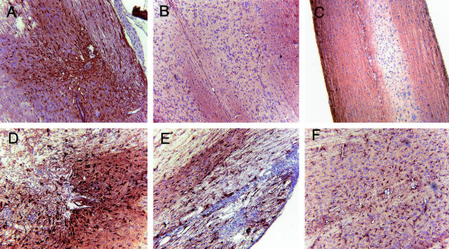
Figure 7.
Gliosis in the spinal cord of T/R+ and T/R− mice. Eight days after SCI, GFAP staining was performed in spinal cord samples of T/R+ (A and B) and T/R− mice (C–F). Section from the site of lesion (A, D, and E) can be observed, as well as sections from the lumbar region (B and F) of the spinal cord. GFAP expression was more abundant and stronger on both injury site and distal areas in T/R− mice than in T/R+. C: The lumbar region of an age-matched T/R− mouse subjected to a mock injury procedure. Note the absence of astrogliosis in this sample. Sections are representative of four animals per group. Original magnifications, ×16.

