Figure 1.
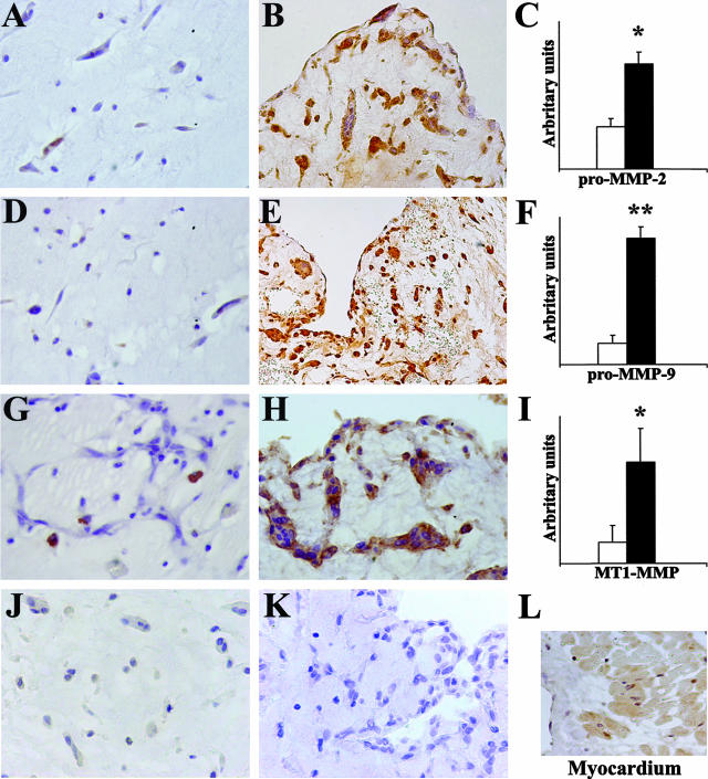
Immunohistochemical evaluation of MMP expression in cardiac myxomas. Embolic myxomas (n = 10) show a high expression of pro-MMP-2, pro-MMP-9, and MT1-MMP (B, E, and H, respectively) whereas in nonembolic myxomas (n = 17) their expression is low or absent (A, D, and G, respectively); TIMP-4 immunostaining is negative in embolic (J) and nonembolic myxomas (K) and positive in control myocardium (L). In C, F, and I, semiquantitative evaluation of immunohistochemical staining expressed in arbitrary units (as reported in Materials and Methods) confirms the higher MMP expression in embolic (▪) compared to nonembolic myxomas (□); results are given as means ± SEM; *P < 0.02 and **P < 0.001 versus nonembolic myxomas. Original magnifications: ×200 (E, L); ×250 (B); ×400 (A, D, G, H, J, K).

