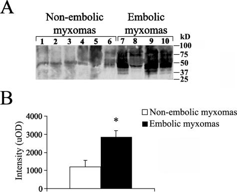
Figure 7.
ECM degradation in cardiac myxoma tissue examined by Western blotting analysis. A: In embolic cardiac myxoma tissues, GAG-containing proteoglycan fragments of less than ∼100 kd are observed and compared to nonembolic tumors. B: Densitometric blot analysis in uOD shows that a greater accumulation of GAG-containing proteoglycan fragments occurs in embolic myxomas. Results are given as means ± SEM; *P < 0.02 versus nonembolic myxomas.

