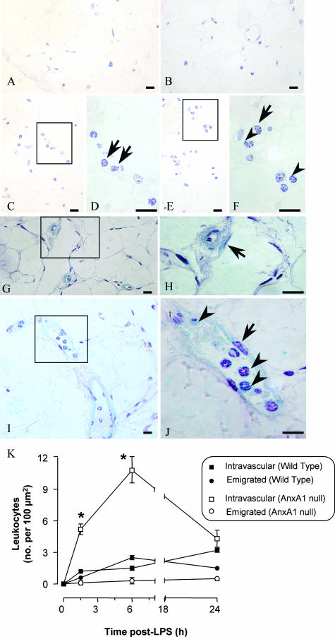
Figure 8.
Leukocyte infiltration into the mesenteric tissue. Wild-type and AnxA1-null mice received 10 mg/kg of LPS intraperitoneally at time 0. At different time points, mesenteries were collected and treated as described in the Materials and Methods (hematoxylin stain). A–F: Tissues from wild-type mice. A and B: Control tissue showing no leukocytes in the connective tissue; C–F: LPS-treated (6 hours) mesenteric tissue showing intravascular (arrows) and transmigrated leukocyte (arrowheads). G–J: AnxA1-null mice. G and H: Control sections with no leukocyte adhesion to the endothelial cells (arrow); I and J: marked degree of cell adhesion (arrowhead) to the endothelial vessels (arrow) as seen after LPS administration (6-hour time point). K: Semiquantitative analysis of the histological sections showing intravascular and extravascular leukocytes in wild-type mice and in AnxA1-null mice. Data are mean ± SEM from five mice per genotype per time point. *P < 0.05 versus corresponding wild-type group values. Scale bars, 10 μm.

