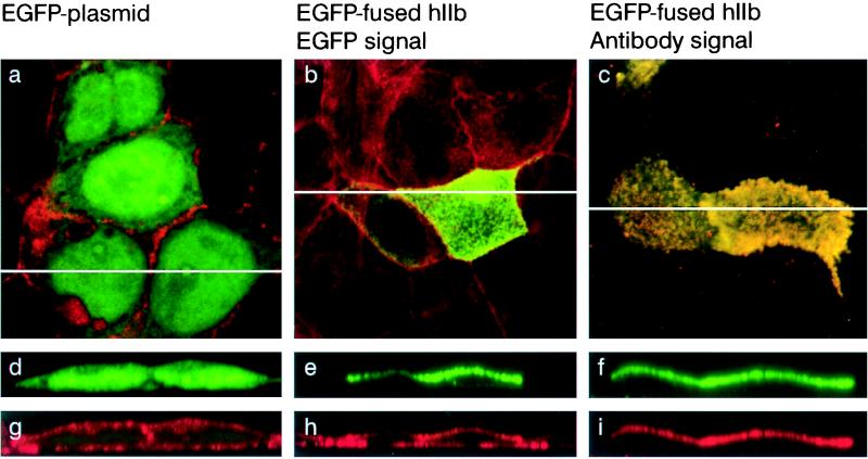
Figure 2.
Expression of hIIb cotransporter in CaCo2 cells. Cells were transfected with EGFP-fused hIIb cotransporter or EGFP plasmid and processed for confocal microscopy. (a–c) Focal planes. (d–i) xy cross-sections. (a, d, and g) Cells transfected with empty plasmid. (b, e, and h) Cells transfected with the EGFP-fused hIIb. (c, f, and i) Cells transfected with EGFP-fused hIIb and stained with anti-hIIb antibody. The endogenous EGFP fluorescence is shown in green and actin and anti-hIIb antibody staining in red. In a–c the fluorescence signals were merged.

