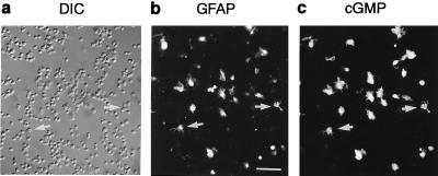
Figure 1.
Location of NO-stimulated cGMP accumulation in cerebellar cell suspension. The same field is shown under differential interference contrast optics (a) and after immunofluorescent staining for glial fibrillary acidic protein (b) and cGMP (c). The cells were fixed after 2-min exposure to DEA/NO (1 μM). Arrows indicate examples of colocalized staining in individual cells. Bar = 50 μm.

