Figure 5.
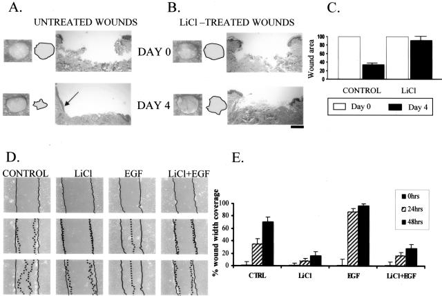
Activation of β-catenin inhibits wound healing and keratinocyte migration. LiCl (ie, nuclear β-catenin) causes delayed wound healing in human skin organ culture wounds. Both gross pictures of untreated (A) and LiCl-treated wounds (B) as well as their histology are shown. Arrow points at epithelial tongue indicating active healing in untreated wounds whereas it is absent in the LiCl-treated wounds. Filled circles indicate the wound surfaces. C: Quantification of the wound size by planimetry shows 70% healing rate of untreated wounds and only 12% healing rate for LiCl-treated wounds. D: LiCl inhibits migration of primary human keratinocytes in wound scratch assay when compared with untreated cells. Inhibition is prominent even in first 24 hours and was further sustained through 48 hours. EGF (positive control) stimulated migration and wound was completely closed after 48 hours. Importantly, this activation of endogenous β-catenin completely blocked EGF-stimulated migration. Full lines indicate initial wound area; dotted lines demarcate migrating front of cells. E: Histograms indicate the average coverage of scratch wounds widths in percent relative to baseline wound width at the day 0 and 24 and 48 hours after LiCl, EGF, and LiCl/EGF treatments.

