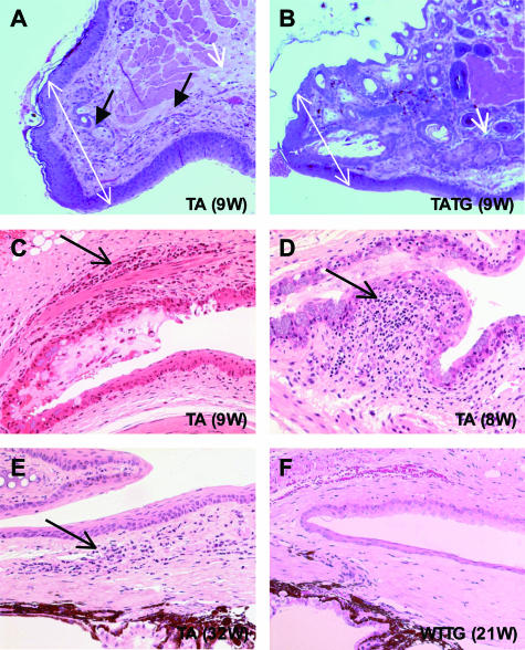
Figure 5.
Photomicrographs showing ocular surface inflammation in Tabby mice. A: Thickened eyelids (double-headed white arrows), absence of meibomian glands (single-headed white arrow), blepharitis (single-headed black arrows), and sparse hair follicles in Tabby eyelids (upper skin). B: Nearly normal eyelids, with partially rescued meibomian gland and fully restored hair follicles (upper skin) in EDA-A1 transgenic Tabby mice. C: Conjunctivitis (arrow) in Tabby. D: Follicular conjunctivitis in the fornix (arrow). E: Limbitis showing inflammatory cells (arrow) infiltrating the junction of the cornea and sclera. F: Normal conjunctiva, limbus, and cornea in transgenic wild-type mice. H&E staining; original magnifications in A and B, ×100; in C to F, ×200.

