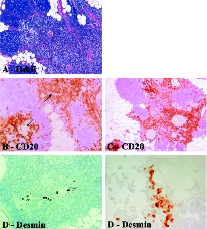
Figure 3.
Photomicrographs showing histopathological characteristics of thymitis. A: H&E staining shows thymus gland with darker cortex (left) and paler medullary region (center and right). Note preserved lobular structure of cortex. B and C: Immunostainings with anti-CD20 antibody reveal B cells (CD20+), which are infiltrating perivascular spaces and surround Hassall’s corpuscles. D: Note Hassall’s corpuscles (center) surrounded by rounded and spindle-shaped myoid cells (desmin+). E: Clusters of desmin-positive myoid cells associated with indentated border within perithymic adipose tissue. Original magnifications: ×10 (A); × 20 (B–D); ×40 (E).

