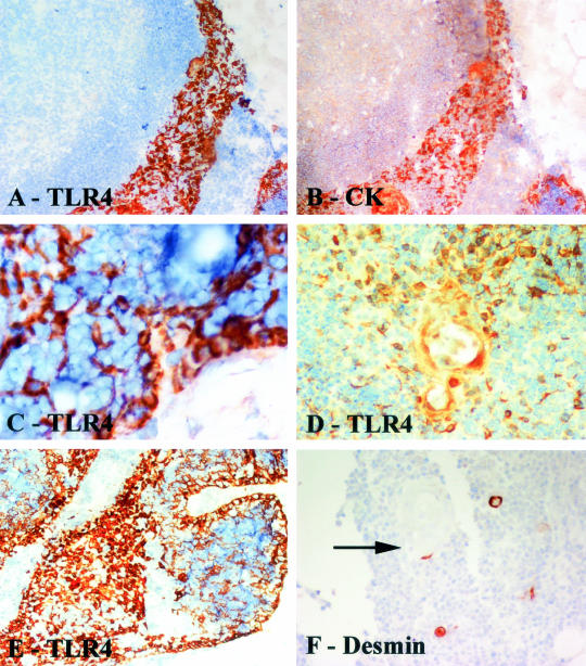
Figure 4.
Immunostaining of thymitis specimens. A and B are serial sections: A, TLR4 immunostaining; B, cytokeratin immunostaining. Note that the TLR4-positive cytokeratin-positive TECs form a band adjacent to the cortex. C: TLR4 stains pericortical epithelial cells interspersed between cortical lymphoid cells. D: TLR4-positive cells clustered around a Hassall’s corpuscle. E: Pericortical and medullary staining of epithelial cells by TLR4. F: Immmunostaining to reveal myoid cells shows that these occasional cells are often associated with Hassall’s corpuscles (arrow). Tissue sections counterstained with hematoxylin. Original magnifications: ×20 (A, B, F); ×40 (C–E).

