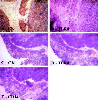
Figure 6.
A, B: Serial sections of thymoma; C–E: healthy young thymus. In A, cytokeratin staining reveals abundant epithelial cells, whereas B show few cells positive for TLR4. In normal thymus cytokeratin immunostaining (C) and macrophage immunostaining (E) reveal that these cells are fairly abundant. D: By contrast TLR4-positive cells are uncommon. Tissue sections counterstained with hematoxylin. Original magnifications: ×20 (A, B); ×10 (C–E).

