Figure 5.
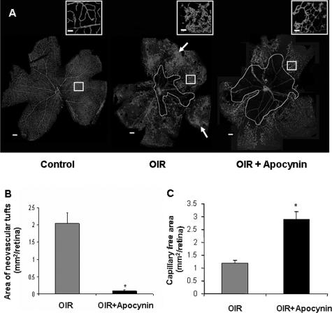
Flat-mounted retinas reacted with GSI lectin to localize neovascularization and capillary obliteration. A: Mice were maintained in hyperoxia from P7 to P12, returned to room air and treated with apocynin (10 mg/kg/day, ip) or vehicle from P12 to P17. Retina from P17 control mouse shows a normal vascular pattern. Retina from nontreated P17 hyperoxia-exposed mouse shows neovascular tufts in the mid-periphery (arrow) and a zone of capillary obliteration around the optic disk. Retina from apocynin-treated P17 mouse shows almost no neovascular tufts and a large capillary-free zone around the optic disk. B: Quantitative comparison of the area occupied by neovascular tufts in the ischemic retinas on P17 shows a significant decrease in neovascularization in the apocynin-treated mice as compared with the nontreated mice (*P < 0.05, n = 5). C: Measurement of the capillary-free zone in the ischemic retinas on P17 shows a significant decrease in revascularization of the central retina of apocynin-treated mice as compared with the nontreated mice (*P < 0.05, n = 5). Scale bars: 100 μm; 20 μm (inset).

