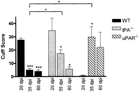
Figure 3.
Perivascular cuff scores for WT, tPA−/−, and uPAR−/− EAE mice. Spinal cords from EAE mice at 17 to 20, 35, and 60 dpi were sectioned longitudinally and stained with an antibody against CD45. Total cuffs were counted in a section area of 4 cm2, and each cuff was given a score according to the degree of infiltration; 1, perivascular inflammation fewer than three or fewer cells deep; 2, more than three cells deep; 3, parenchymal infiltrate. A total of three slides from different mice were counted per time point and the data are shown as the mean ± SEM; *P < 0.05 and ***P < 0.001, significance illustrated versus control unless illustrated by a bar. Cuff counts (not illustrated) showed a similar pattern as the cuff scores.

