Figure 5.
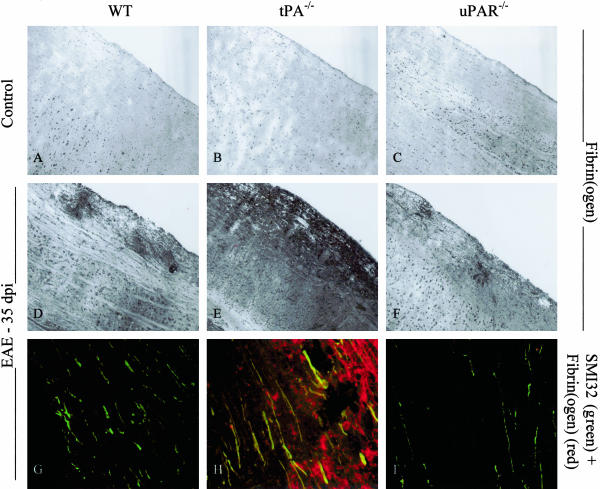
Fibrin(ogen) localization in EAE. i: Spinal cords were removed from control animals and mice 35 dpi onwards of EAE and cut longitudinally. Frozen sections were stained with an antibody against fibrin(ogen), and double-fluorescence staining was performed with antibodies against fibrin(ogen) and SMI32, a marker of nonphosphorylated neurofilament. Fibrin(ogen) can be seen surrounding perivascular cuffs in sections from EAE animals (D–F), but in much greater amounts in tPA−/− mice. Co-localization of fibrin(ogen) on SMI32-positive axons is seen in sections from tPA−/− EAE mice (H) but not in sections from WT or uPAR−/− animals (G or I). Original magnifications: ×100 (A–F); ×400 (G–I).

