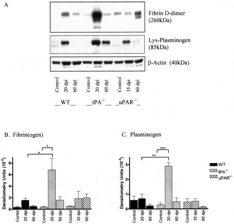
Figure 6.
Western blotting of fibrin(ogen) and plasminogen. Spinal cords from control and EAE mice were homogenized for protein extraction. Levels of fibrin(ogen) and plasminogen were detected by Western blotting and were quantitatively measured by densitometry scanning (A) and results are shown as arbitrary densitometry units ± SEM. Blots were reprobed with anti-actin to ensure equal loading of proteins. Levels of fibrin(ogen) (B) and plasminogen (C) were significantly increased during the acute phase of EAE in tPA−/− mice when compared to control mice and WT mice at the same stage of disease. *P < 0.05, **P < 0.01, ***P < 0.001.

