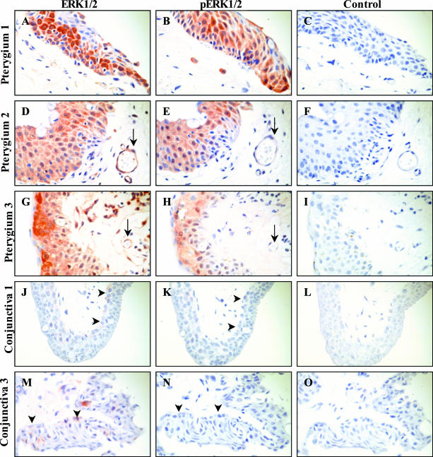
Figure 3.
Localization of ERK1/2 in pterygia. Pterygia (A–I) and normal conjunctiva (J–O) were serially sectioned and stained for total ERK (A, D, G, J, and M) or pERK (B, E, H, K, and N). Control reactions included omitting the primary antibody (C and F) or applying a relevant isotype control IgG antibody (I, L, and O). Tissue sections in A to C and J to L were derived from one subject and tissue in G to I and M to O were derived from another patient. Pterygium tissue in D to F was obtained from a third donor. Red staining denoted positive immunoreactivity. Arrows in D and G indicate a positively stained blood vessel. Whereas the arrows in E and H point to the same unstained vessel. Arrowheads in J and M illustrate mild reactivity for total ERK in the basal conjunctival epithelium, whereas the arrowheads in K and N indicate the absence of any reactivity for phospho ERK (pERK) in the same region of the conjunctiva. This pattern of expression was representative of all diseased and normal tissue specimens analyzed. Original magnifications, ×500.

