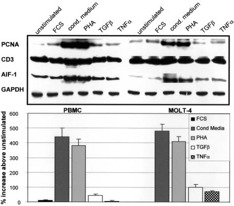
Figure 2.
Expression of AIF-1 protein in activated human lymphocytes. Human PBMCs and the human T-myeloblast cell line, MOLT-4, were serum-starved for 48 hours, and protein from 1) serum-starved, and cells treated for 40 hours with 2) 15% fetal calf serum; 3) T-cell conditioned media; 4) PHA; 5) transforming growth factor-β; and 6) tumor necrosis factor-α. Extracts from these cells were subjected to Western blot with anti-AIF-1, proliferating cell nuclear antigen, CD3, and GAPDH antibody. Bands were quantitated by densitometry, and normalized to GAPDH. Blot shown is representative of three performed from different groups of PMBC and MOLT-4 cells. Bar graph indicates percent expression above unstimulated cells for three groups of experiments.

