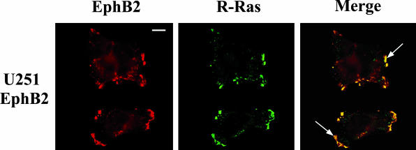
Figure 3.
Co-localization of EphB2 and R-Ras in glioma cells. U251 cells stably transfected with EphB2 were co-stained for R-Ras (fluorescein-isothiocyanate stained) and EphB2 (Cy3 stained), and examined by confocal laser microscopy. Arrow in panel merge indicates that R-Ras co-localized with EphB2 in lamellipodial structure. Scale bar, 10 μm.

