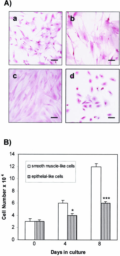
Figure 2.
Morphological appearance and growth rate of the two cell types isolated from the TSC human angiomyolipoma. A: H&E staining of flat-elongated smooth muscle-like cells at different magnifications (a, b) and at confluence (c), and rounder epithelial-like cells (d). B: Growth rate of smooth muscle-like and epithelial-like cells 4 and 8 days after plating. *P < 0.05 and ***P < 0.001 indicate significant differences versus smooth muscle-like cell proliferation. Scale bars: 40 μm (a, d); 20 μm (b, c).

