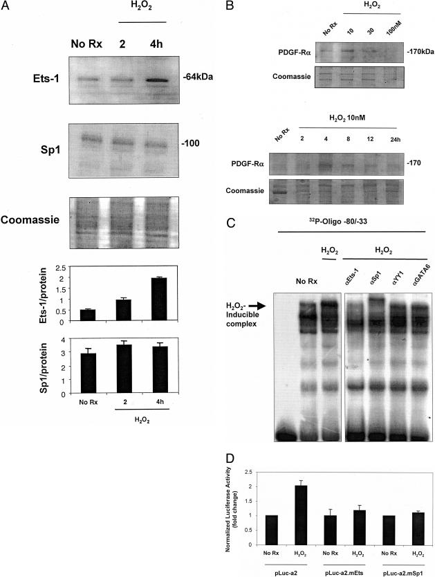
Figure 7.
H2O2 induces Ets-1 protein expression and PDGF-Rα transcription via the −45TTCC−42 motif in the proximal promoter. A: Growth quiescent cells were incubated with 10 nmol/L H2O2 for 2 and 4 hours. Ets-1 and Sp1 protein levels were assessed by Western immunoblot analysis (20 μg total protein for Ets-1 analysis and 10 μg for Sp1). Ets-1 protein levels increased compared to basal levels with addition of 10 nmol/L H2O2 compared to untreated cells. Sp1 protein levels were unchanged compared to basal levels with addition of 10 nmol/L H2O2 compared to untreated cells. Coomassie-stained gel demonstrates unbiased loading. Densitometric analysis was performed on bands of interest and relative to Coomassie-stained protein. B: SMCs were incubated with various concentrations of H2O2 for different times and PDGF-Rα protein levels assessed by Western immunoblot analysis (10 μg of total protein). Coomassie-stained gels demonstrate unbiased loading. C: EMSA using nuclear extracts of SMCs that had been incubated with 10 nmol/L H2O2 for 4 hours. The H2O2-inducible nucleoprotein complex was eliminated by antibodies to Ets-1 and Sp1, but not YY1 or GATA6. D: SMCs were transfected with 10 μg of either pLuc-a2, pLuc-a2.mEts, or pLuc-a2.mSp1 for 24 hours followed by the addition of 10 nmol/L H2O2 for 24 hours. Firefly luciferase was determined in cell lysates after 24 hours. The y axis represents the fold change in PDGF-Rα promoter activity. The result is representative of at least two independent observations.

