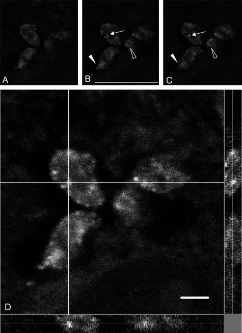
Figure 2.
Confocal microscopy observations of intraparasitic TGF-β immunoreactivity. A to C correspond to three of nine successive confocal sections taken around the middle of the z axis. Staining is observed in cytoplasmic granules (arrows) and in the flagellar pocket (arrowheads) both in longitudinal (filled arrowheads) or in sagittal (open arrowheads) sections of the parasites. D: Large magnification of a confocal single-plane image of TGF-β detection in intracellular T. cruzi. The analysis was performed along the x-y axes (central), the x-z axes (bottom), and the y-z axes (right). The white lines indicate the axes along which the deconvolution was performed. Note the fluorescent internal vesicles clearly visible along the z axis. Scale bars, 10 μm.

