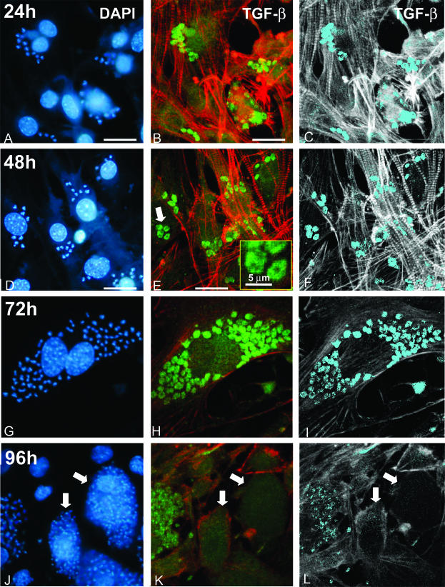
Figure 5.
Parasite life cycle-dependent immunostaining of TGF-β in intracellular forms of T. cruzi. Cultured cardiomyocytes were infected by T. cruzi trypomastigotes and the localization of TGF-β immunoreactivity was analyzed after 24 hours (A–C), 48 hours (D–F), 72 hours (G–I), or 96 hours (J–L) of infection by triple labeling of DNA with DAPI (A, D, G, J), TGF-β with anti-TGF-β-FITC complexes, and actin fibers with phalloidin-TRITC (green and red, respectively, in B, E, H, K). C, F, I, and L correspond to image-processed views from the original confocal images shown in B, E, H, and K, to stress (in light blue) the localization and the progressive decay of TGF-β immunoreactivity in the parasites. The inset in E shows a larger magnification of the parasites pointed out by the large arrow. In J, K, and L, the arrows show two cells containing a large amount of trypomastigotes that are clearly poorly immunoreactive for TGF-β. Scale bars, 20 μm (unless otherwise indicated).

