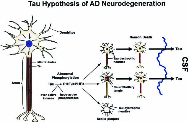
Figure 1.
The misfolding, fibrillization, and sequestration of tau into filamentous inclusions is schematically depicted and is described in greater detail in the text. Proceeding from the normal neuron on the left, this tau hypothesis of AD neurodegeneration predicts that the cascade of events depicted schematically will compromise the function and viability of neurons (shown to the right) through the pathological conversion of normal tau into PHFtau, which forms NFTs and dystrophic tau neurites thereby depleting levels of functional MT-binding/stabilizing tau below a critical point that results in the depolymerization of MTs and a disruption of axonal transport. As shown on the far right, the dissolution of degenerating tangle and dystrophic neurite-bearing neurons in the AD brain releases abnormal tau into the extracellular space resulting in elevated levels of cerebrospinal fluid (CSF) tau, which is one of the most robust biomarkers of AD in living patients.

