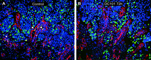
Figure 4.
Endothelial proliferation within intratumoral stromal strands. Fluorescence micrographs showing proliferating endothelium within intratumoral stromal strands of control (A) and 72-hour DC101-treated (B) sections. Triple staining of Hoechst nuclear staining (blue), collagen IV (red), and BrdU (green) revealed tumor areas (predominantly blue staining tumor nuclei) interspersed with collagen IV (red) staining endothelial basement membrane (luminal structures) and tumor epithelial basement membrane (red, stromal-tumor interface). Proliferating nuclei (green) could be seen within both tumor and stromal areas. BrdU staining (green) of collagen IV-positive luminal areas (red) revealed proliferating endothelial cells (A, open arrowheads). A: Control 24 hours; B: 72 hours DC101. Original magnifications, ×200.

