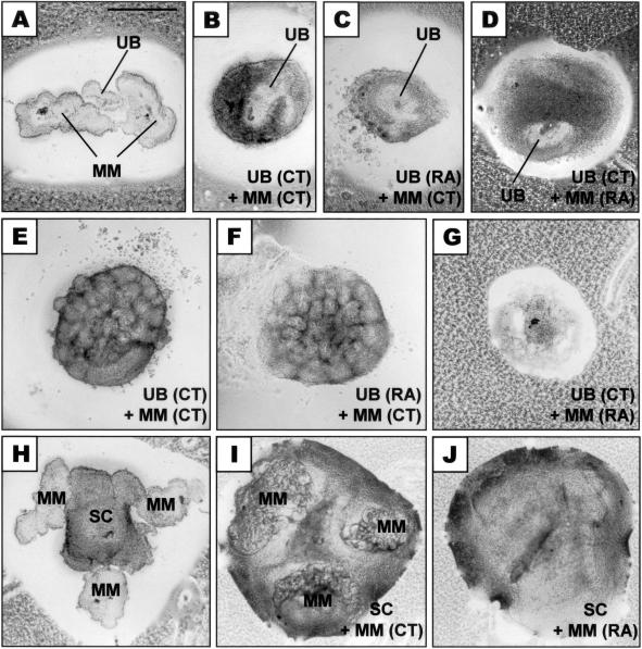
Figure 2.
Development of recombinants in culture. A–G: Co-culture of two pieces of MM with one piece of UB isolated from metanephroi of control (CT) or RA-exposed embryos (RA). Recombinants before (A), or after 2 days (B–D) or 7 days (E–G) in culture. Note that RA-treated UB when recombined with control MM (C and F) showed a similar pattern of growth and differentiation as recombinants between control UB and control MM (B and E). However, MM from embryos exposed to RA could not respond to control UB and degenerated (D and G). H–J: Co-culture of MM with embryonic spinal cord (SC). At the beginning of culture, three pieces of MM were placed in close proximity to one piece of SC (H). After 7 days in culture, while SC could induce tubulogenesis of control MM (I), MM from RA-exposed embryos could not respond to SC and degenerated (J). Scale bar, 0.5 mm.

